e-Stroke Suite 10.0 Provides First-Ever Clot Detection from Non-Contrast CT Scans, a Paradigm Shift Enabling Clinicians to More Broadly Identify Thrombectomy-Eligible Stroke Patients.
Oxford, UK, 16th September 2020
Brainomix have announced the launch of its e-Stroke Suite 10.0, the latest version of the company’s award-winning stroke imaging platform. The new release incorporates a series of revolutionary AI technologies, including:
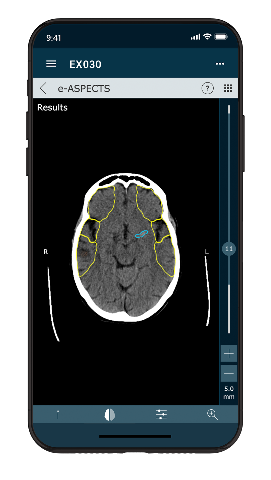
- Detection and measurement of both Large Vessel Occlusion (LVO) and hyperdense volumes (which may indicate bleeding) from non-contrast CT scans with e-ASPECTS;
- Graphical visualization of CTA acquisition timing, arch-to-vertex CTA viewing, and the capability to derive CT angiography results from a CT perfusion scan with the enhanced e-CTA module;
- A new feature enabling clinicians to enter patient data (such as NIHSS, last known well time, etc.) that can then be shared via the e-Stroke Cloud and e-Stroke Mobile app;
- A “virtual hub” enabling secure real-time sharing of images and patient characteristics across a previously unconnected network, addressing another barrier to expedient patient transfer between many hospitals.
“At Brainomix we are focused on advancing the value of simple imaging, and with these new ground-breaking capabilities, our aim is to empower physicians to make more confident decisions in a timely manner, expanding patient access to life-saving treatments,” noted Eric Greveson, Chief Technology Officer at Brainomix. “We are proud of this latest release, which builds on our legacy of continual innovation, while also firmly establishing the e-Stroke Suite as the most comprehensive stroke imaging solution available.”
The detection of LVO – one of the leading determinants of patient eligibility for mechanical thrombectomy – from universally available NCCT scans may have profound clinical value, enabling earlier decisions about treatment and formal CT angiography if not routinely acquired. As noted by Dr Christian Herweh, Consultant in the Department of Neuroradiology at Heidelberg University Hospital: “With the e-Stroke Suite now enabling automated detection of hyperdense artery sign suggestive of LVO on NCCT, more patients should be flagged as thrombectomy eligible – particularly those admitted first to smaller hospitals with less established stroke pathways, who can then be transferred for treatment to a specialist center.”
Prof Simon Nagel, Managing Senior Consultant in the Department of Neurology, also spoke of the clinical benefits associated with the new software: “The e-Stroke Suite is now able to differentiate between hemorrhagic and ischemic stroke by detecting hyperdense structures in the brain, and the e-ASPECTS module can quantify the extent of ischemic hypodense regions by the ASPECTS score and volumetrically, providing our team with the most complete AI-powered imaging application for NCCT in the AIS pathway.”
About Brainomix
Brainomix is an Oxford-based company specializing in the creation of AI-powered software solutions to unlock the potential of life-saving treatments. Founded in 2010 as an offshoot of the University of Oxford, its e-Stroke Suite provides clinicians with the most comprehensive stroke imaging solution, helping to interpret images and facilitating faster, more confident treatment decisions for patients with suspected stroke. To learn more about Brainomix and its technology visit www.brainomix.com, and follow us on Twitter, Facebook and LinkedIn.


