Vision to Precision

Brainomix 360 e-Lung

Brainomix e-Lung is an FDA-cleared, CE-marked, AI-powered imaging software platform that automatically identifies and quantifies features on CT lung scans using a configured clinical workflow. Quantification of CT features can enable clinicians to more easily identify visual changes over multiple timepoints.

Request a demo
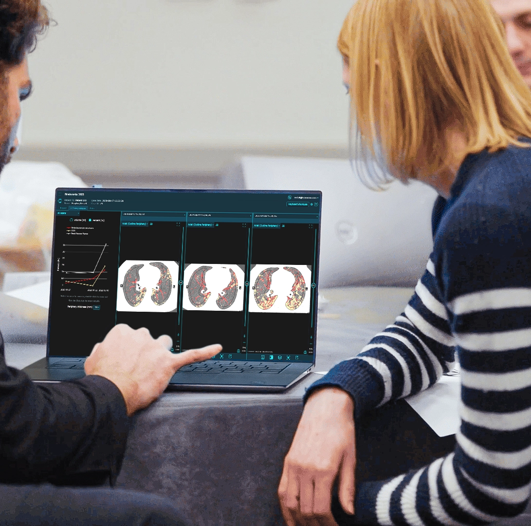
Brainomix 360 e-Lung laptop interface
The Challenge of Earlier Detection
Diagnostic Difficulties
Patients with ILD can develop Progressive Pulmonary Fibrosis (PPF), which causes irreversible lung damage and leads to early mortality. The key to the best outcome and survival for patients is early initiation of treatment. However, identifying patients eligible for treatment based on imaging can be challenging, even for experts.

Doctor discussing medical information on a computer with an elderly couple in a bright office.
INBUILD clinical trial CT analysis on laptop
INBUILD LANDMARK TRIAL

Validated by the INBUILD Trial

Through a research collaboration and partnership with Boehringer Ingelheim, the global leader in pulmonary fibrosis therapies, Brainomix were granted privileged access to the landmark INBUILD clinical trial dataset to run the first quantitative image analysis.

The analysis used quantitative metrics from CT images. The authors were able to accurately and sensitively facilitate identification of PPF, including the assessment of PPF progression.

*This study has not been evaluated by FDA.

Doctors reviewing CT scans
Lung CT scan visualization
Revise PPF Study

Researchers Demonstrate Earlier Detection of PPF in New Study

Results from a retrospective study with the University of Chicago, Weill Cornell Medicine, and the University of Alabama at Birmingham were presented at ERS and CHEST. The authors showed:

They could identify CT progression in 74% of patients deemed clinically stable.

They accurately identified patients at risk of developing future PPF from a single baseline scan.

*This study has not been evaluated by FDA.

“The data we have shown for e-Lung is very promising, and the ability to objectively assess parenchymal changes to predict disease trajectory and treatment responses could really help us personalize treatment decisions and improve outcomes for patients living with pulmonary fibrosis.”

Dr Teja Kulkarni

University of Alabama at Birmingham

*This testimonial has not been evaluated by FDA.

E-LUNG READER STUDY

e-Lung Featured in a New Reader Study

A new reader study was presented at ECR 2026 by Dr Logan Sun (Royal Brompton Hospital, London). Five readers, blinded to clinical data, independently reviewed serial CTs side-by-side from 102 patients with non-IPF fibrotic ILD. All patients in this cohort demonstrated marginal FVC decline of 5 - 10%.

Readers categorized each case as either stable or progressive disease based on visually estimated changes in ILD extent.

Overlays configured to quantitatively visualize parenchymal radiological features were applied.

In cases initially categorized as stable, readers could either retain the original categorization or change to progressive. The quantitative CT imaging features were used to identify 22 to 40 cases per reader for re-evaluation. Readers changed PPF categorization in 45 - 94% of these cases, demonstrating improved reader performance.

e-Lung at ECR 2026 conference

*This study has not been evaluated by FDA.

Ready to transform lung care?

Discover how you can integrate e-Lung into your Lung CT clinical workflows.