



Stroke Visionaries: Dr. Achala Vagal
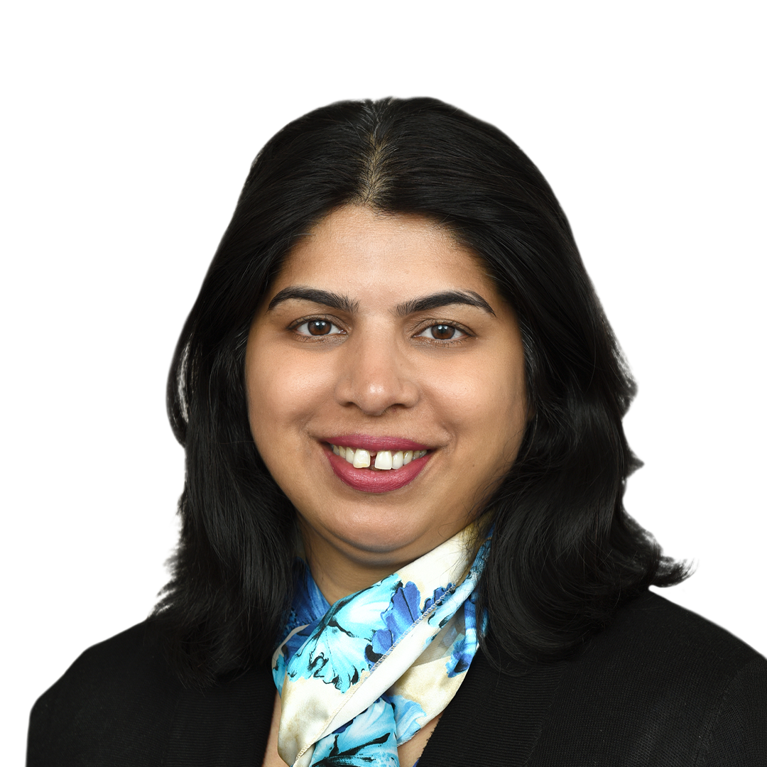
As part of our interview series "Stroke Visionaries," we had the pleasure of meeting with Dr. Achala Vagal.
Dr. Achala Vagal is the Chair of the Department of Radiology at the University of Cincinnati College of Medicine, where she also serves as Associate Dean of Clinical and Translational Science and Training. A tenured Professor of Neuroradiology, she earned her medical degree from Grant Medical College and Sir J.J. Group of Hospitals in Mumbai, India, completed her diagnostic radiology residency at the University of Mumbai, and went on to fellowships in neuroradiology and imaging at the University of Cincinnati, followed by a master's degree in clinical and translational research. She is board certified by the American Board of Radiology in both diagnostic radiology and neuroradiology. Dr. Vagal is a nationally and internationally recognised expert in neuroradiology and stroke imaging. She directs the National Imaging Management Center for NIH StrokeNet, leading the imaging core lab for large multicentre national and international clinical trials, and is co-director of the Center for Clinical and Translational Science and Training (CCTST) at University of Cincinnati. Her research focuses on stroke imaging biomarkers and brain health across clinical trials and population studies, and she has a particular interest in the thoughtful clinical integration of AI in radiology.
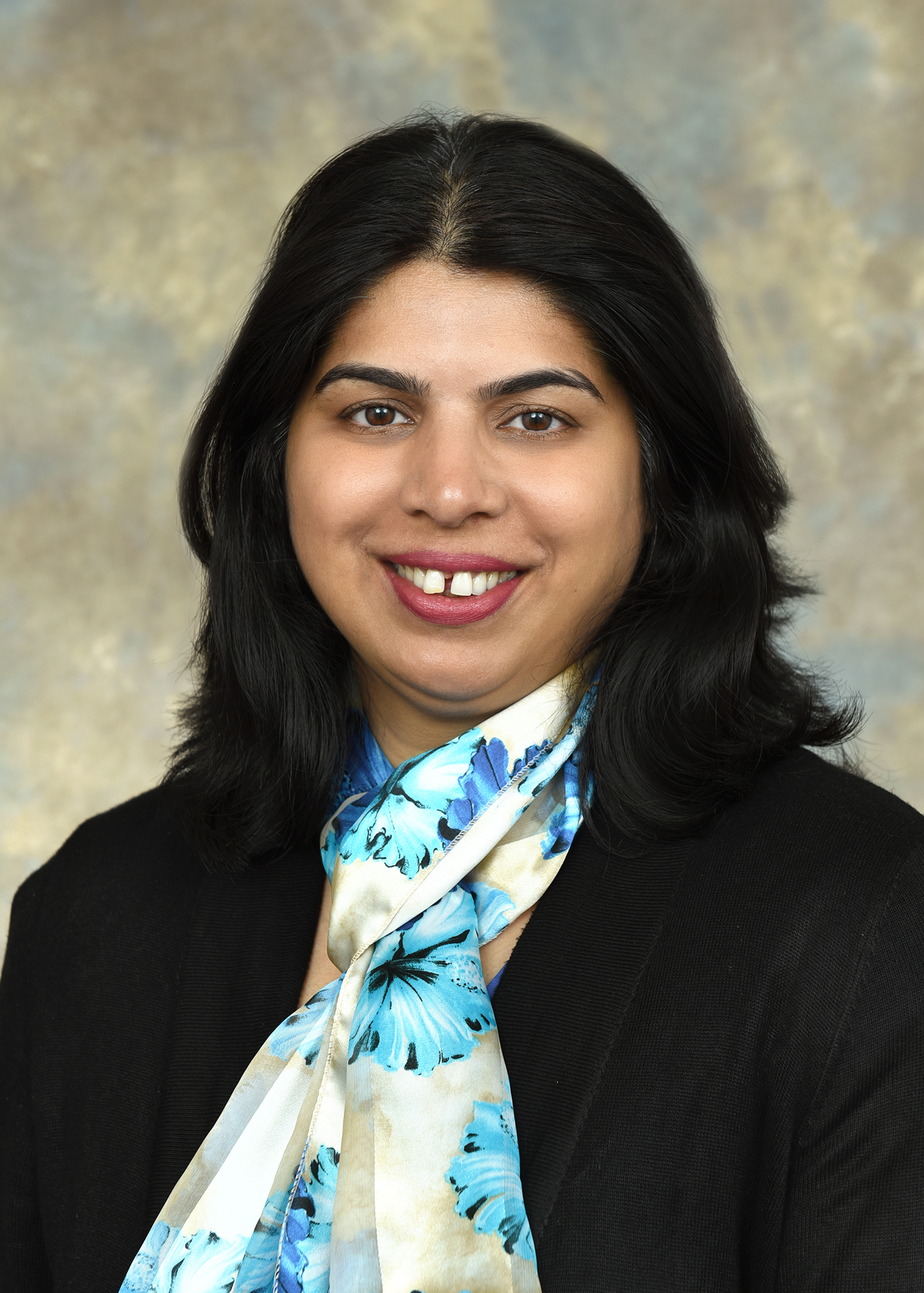
Thank you so much for joining us on this interview series! Can you talk me through your career path? Why medicine, and what moved you towards radiology and then stroke?
I'm originally from India, so I trained and did my residency there. But I always had this dream that I should come and learn from the experts, the people who have actually written the books in radiology. Then I got the opportunity to come to Cincinnati, and that was it. I was living my dream, learning from the best radiologists in a department with a very rich culture of education.
The stroke piece was very organic. One of my mentors, Dr. Thomas Tomsick, a pioneer in stroke treatment, said to me one day, you should do stroke work. I said, I'm not an interventionalist, I'm a diagnostic radiologist. He said there is a need for diagnostic neuroradiologists in stroke, and I think you should do it. He connected me with a few other people in Cincinnati, Drs. Broderick, Pancioli, Khatri, and I started on that journey: imaging core lab work and clinical trials. One thing led to another, and here I am after so many years, still doing stroke research.
And back in India, how did you decide on medicine, and then radiology?
I have wanted to be a doctor since I was five. I don't know how or why, but I just knew. My journey to radiology is not a wildly inspiring story, but it's a true one. I was actually going to be an OB/GYN, that's what I wanted my entire life. Then somebody in Mumbai suggested radiology. I said, what's that? They told me it was going to be the next big thing. So I applied, I got in, and I still wasn't sure. Then I realised the radiology department was the only centrally air-conditioned department in the Mumbai heat. So I thought, this is it. It wasn't for the love of radiology, it was the love of air conditioning.
But I have always maintained that things happen organically, things work out for you, but in the end it is the person who brings joy to the work. It doesn't go the other way around. That's my biggest advice to trainees. There is no perfect job, no perfect career. No job, no field is ever going to give you joy if you don't bring it.
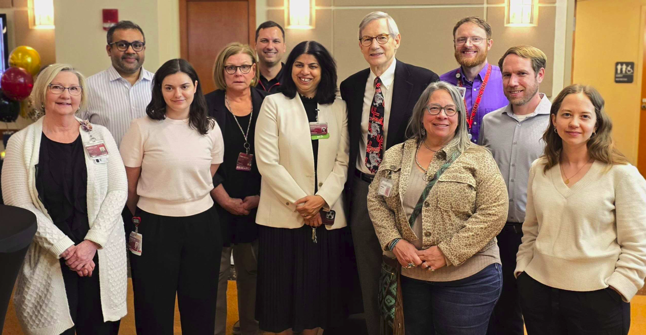
You're now chair of radiology, and you've talked about making Cincinnati a pioneer for AI. Where did that vision stem from?
Becoming chair of the department where I came as a trainee, to learn from the masters, and now to serve in a different role, is very important to me. Radiology has always been the pioneer when it comes to AI, because imaging has digital data, and that data is gold for AI. What I want to do in our department is the clinical implementation of AI, but in a thoughtful way: integrate it, make it practical, and when you deploy it, if things don't work well, you should be able to trace it back.
The biggest problem in radiology worldwide, including the US, is a huge workforce shortage. I believe in augmented intelligence, and it will help us. But it will take time, on the side of the AI companies and the radiologists, to become good partners, and to implement it in a way that is user friendly. The end result should be better reports, more efficient work, and better care for patients. It's that in-between, the clinical implementation, where there are still so many challenges.
What is a challenge you'd like to overcome when it comes to clinical implementation of AI?
There are two big ones. First, AI sometimes over-promises and sometimes under-delivers on accuracy, sensitivity and specificity for imaging findings.
The second is that it's hard to show a clear return on investment for the administrators. AI is getting more and more expensive. Every module, every algorithm, is one more price tag. So what is the ROI? Is it going to make us faster? Often, no. Are we going to hire fewer radiologists? No, we still have all the work. When hospital margins are reducing and finances are tight, having a business ROI is very difficult.
Stroke is different, because stroke was one of the pioneers in AI very early on. It changed patient care, it was a paradigm shift. Some lessons can be learned from stroke and applied to other parts of radiology. Stroke is way ahead.
Are there particular cases where AI has been a valuable partner?
One of the biggest is the vessel occlusion algorithm. For the AI to flag a vessel occlusion before the radiologist has even seen it, particularly in rural settings where there may not be 24/7 or specialised radiologist coverage, that ability to help with patient triage is huge. There are not enough radiologists to go around every rural part of the world. So I've seen that work well, where patients could be triaged even before a radiologist read the scan.
I've also seen the AI miss things, and people who aren't comfortable with the imaging, or don't understand the algorithm, may not catch it. A classic example: initially, modules were trained only for ICA and M1 vessels, but they were marketed as large vessel occlusion detection. The posterior circulation, like the basilar, was often not included. So the algorithm may or may not pick up a basilar occlusion. That's not the fault of the AI. If it was never trained for it, it's not going to pick it up. It's like any other tool. If you don't understand how it was trained and what it can and cannot do, then it's not a good partner. AI is augmented intelligence. It's going to augment the workflow of physicians, but for that, you have to understand the tool.
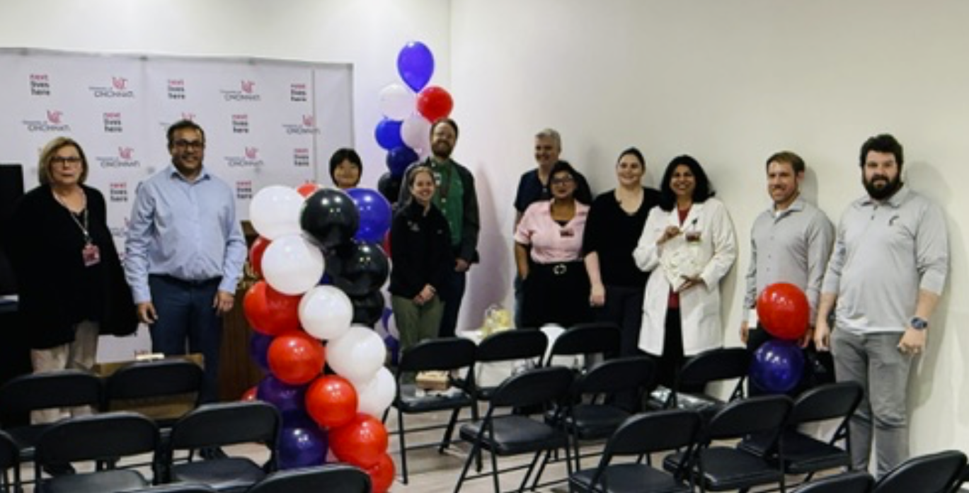
So an important key in that case is knowing the limitations of the tool you're using?
Know the limitations, but also know how the tool has been trained. If I know what kind of data was used, I can make an informed decision. The other big thing is that you have to work closely with industry. Ask them, show me the white paper, share how this was built. My experience is that the industry is willing to share, because the end goal is better patient care.
Radiologists are the imaging experts. So it almost behooves us to work closely, whether with academic or industry partners, then we inform how the AI is trained, and we educate the non-radiologists on the pros and pitfalls. We are the experts at the end.
And working closely with industry lets you shape the tools you'll need in future.
Absolutely, because we understand the needs, and where science is moving. More and more information can be gleaned from the images. I always say an image is worth a thousand words. That image has so much information that can be extracted. But the AI also needs to be explainable. If it extracts information I cannot understand, then the output may or may not be clinically significant. So it's about making it explainable, and understanding where the limitations and pitfalls are.
Alongside your many other roles, are you still involved in research?
I still do research. I run the Imaging Management System for NIH StrokeNet, so we run the imaging core labs for multiple clinical trials. That covers coordinating the study sites for imaging collection according to each protocol, making sure everything is properly anonymised, tracking the imaging data transfers, and running quality control on the imaging data, through to the imaging analysis itself and supporting the central readers.
I get to work with the thought leaders in the field and stay close to what's going on in the stroke clinical trial space. I'm also a Co-Director and multi PI for our Center for Clinical and Translational Research and Science, where we build the infrastructure for research, not just for one project or grant, but the processes and infrastructure so that investigators from any field can use it.
I've become busy, but this is an important part of giving back to radiology, imaging and stroke. What I like to remind myself and my team is that we are a brick in the cathedral of stroke care, and we want to continue to do that. The platforms, the processes, the infrastructure, so the work can go on way beyond one investigator, one discovery, one institution. That's the goal.
What do you like to do outside of work? What gives you joy?
I wouldn't call it a hobby, but I'm an ardent student of yoga and meditation. I'm a certified yoga teacher, so I like to get on the mat and meditate. That's the one thing that keeps me grounded and calm. It's the thing from which everything else comes, including the joy in work and in life. That's my go-to space.
If you or someone you know is driving change in stroke care, we’d love to hear from you. Please send an email with your nomination to Emma Houtz at ehoutz@brainomix.com. Stay tuned for more editions of Stroke Visionaries.
Ready to transform stroke care?
Join 400+ hospitals worldwide already using Brainomix 360 Stroke to save more lives, faster.

Brainomix Limited is a company registered in England and Wales at OX2 0JJ. Company Registration No. 07426406


Brainomix © 2026. All rights reserved